
Longitudinal DNA samples from peripheral blood
Longitudinal peripheral blood DNA samples were obtained from blood serum samples biobanked as part of the UKCTOCS (ISRCTN22488978; ClinicalTrials.gov identifier: NCT00058032). UKCTOCS is a multicentre randomized controlled trial which recruited 202,638 postmenopausal women aged 50 to 74 years, in England, Wales and Northern Ireland between 2001 and 2005, to assess the impact of screening on ovarian cancer mortality.3. UKCTOCS eligibility criteria and trial details have been described previously58. UKCTOCS was reviewed and approved by the North West Multicentre Research Ethics Committee (REC reference 00/8/34) and participants provided written, informed consent for the use of their samples and data in ethically approved secondary studies. Our study, using UKCTOCS samples and data, was reviewed and approved by the South Central Berkshire B Research Ethics Committee (REC reference 18/SC/0481). This study was carried out in accordance with all applicable guidelines and regulations.
Upon enrollment in the UKCTOCS, 50,640 participants were randomized into a “multimodal screening group”, which involved annual blood serum samples for up to 11 years, until December 31, 2011. All samples were collected using a standardized protocol. Blood was collected in gel tubes (8 mL gel separation serum tubes; Greiner Bio-One 455071) at the testing centers and transported daily at room temperature to a central laboratory, where the samples were centrifuged (1,500g for 10 min) and the separated serum aliquoted into 10 × 500 µl straws using a semi-automated MAPI (IMV) platform. Straws were stored in liquid nitrogen tanks at the central laboratory which, once full, were transported to an HTA-approved commercial cryofacility (Fisher Bioservices, UK, until March 2018, NIHR Biocentre from April 2018 to July 2023 and BioDock since then). The median time from sample collection to centrifugation (approximately 22.1 hours) resulted in leakage of leukocyte DNA into serum.59. Health outcomes of UKCTOCS participants were tracked via linkage to national cancer, death and administrative registers. In 2017, 47 women in the “multimodal screening arm” had incidentally developed AML (ICD-10 C92.0) and had a sufficient serum sample at least two time points before AML diagnosis. These 47 women were diagnosed with AML at a median age of 72 years (range 53–83 years, interquartile range 8 years) and serum samples were biobanked from a median of 5 time points (range 2–10, interquartile range 3.5) before AML diagnosis (Extended data, Fig. 1a,b). We obtained 1 ml of serum from each of the available annual time points (249 samples) from these 47 women (“pre-AML cases”), as well as from serial samples corresponding to time points from 47 age-matched UKCTOCS participants who remained free of blood cancer (“controls”). A control was then excluded due to the development of MDS. No statistical method was used to predetermine the sample size; all eligible pre-AML cases were included. Randomization was not applicable because this was an observational study and investigators were not blinded to case-control status during sequencing and analysis. DNA was extracted by LGC Genomics, using an adapted Kleargene method with magnetic beads on KingFisher 96, and eluted in 10 mM Tris-Cl, pH 8.0. DNA was quantified using PicoGreen, giving an average of 279 ng ml-−1 DNA (range 11-814 ng ml−1 serum) in pre-AML samples and 270 ng ml−1 DNA (range 11 to 1313 ng ml−1 serum) in control samples (extended data, Figure 1c).
Information on any diagnosis of MDS (ICD-10 code D46 or C94.6), CMML (ICD-10 code C93.1), or MPN (ICD-10 codes D45, D47.1, D47.3, D47.4, C92.1, C92.2, C94.4) or other hematologic malignancies (e.g., lymphoma or myeloma) was obtained for all pre-AML cases. cases and controls via record linkage with the National Cancer Registry (England/Wales), Hospital Episode Statistics (HES) or Patient Episode Database for Wales (PEDW), Office for National Statistics mortality records and, for participants in Northern Ireland, the Central Services Agency and the Northern Ireland Cancer Registry. Information on diagnoses of non-haematological malignancies was also obtained via links to national cancer registries, HES/PEDW databases and Office for National Statistics mortality registers. These clinical diagnoses were extracted to characterize myeloid disease histories, prior malignancies, and treatment exposures that provide relevant clinical context for secondary and treatment-related AML settings (see Supplementary Note 5 for detailed case annotations). When a diagnosis appeared in more than one data source, the first recorded entry was considered the diagnosis date. Recorded diagnosis dates therefore reflect the first appearance of a diagnosis in national registers and may not correspond to the biological onset of the disease. Potential coding errors were assessed by triangulation between available sources: diagnostic entries that appeared only once in HES/PEDW and were discordant with other HES/PEDW episodes, cancer registry data, or death certificate information were treated as probably miscoded and excluded. Information on receipt of chemotherapy (ICD-10 codes Z51.1, Z51.2, Z92.6) or radiotherapy (ICD-10 codes Z51.0, Z92.3) before AML diagnosis (or, for controls, before age-matched case at AML diagnosis) was obtained from the HES and PEDW databases. For participants with a record of chemotherapy, the indication was determined by cross-referencing diagnosis codes in HES/PEDW and/or cancer registry data. Chemotherapy codes temporally associated with AML diagnosis and lacking evidence of prior malignancy were considered AML-directed and were not classified as pre-AML exposure. No pre-AML cases or controls were recorded as having previously received radiotherapy. Information on smoking, where available, was obtained from UKCTOCS participant questionnaires and HES databases (ICD-10 code Z72.0). The latest updates from all linkage sources were received in 2020. As HES and PEDW only cover NHS-funded care in England and Wales, it is possible that diagnoses or treatments delivered privately, overseas or elsewhere in the UK may not have been captured. Supplementary Table 10 summarizes the timing of any recorded diagnosis of MDS, CMML, MPN, or other hematologic or non-hematologic malignancy, as well as prior exposure to chemotherapy and smoking history, for all pre-AML cases and controls, with mortality outcomes (cause of death and survival interval) summarized for pre-AML cases only.
Cell type deconvolution
To estimate the proportions of different types of blood cells (B cells, natural killer cells, CD4+ T cells, CD8+ T cells, monocytes, neutrophils and eosinophils) present in the samples, we used the R package EpiDISH with the reference dataset ‘centDHSbloodDMC.m’60. Whole-genome bisulfite libraries were prepared for 94 UKCTOCS samples from 23 pre-AML cases and 23 controls, some of whom contributed at more than one longitudinal time point. Of these 94 samples, 88 met the minimum coverage criterion (at least 286 of 333 CpGs covered) and were included in the deconvolution analysis (average depth 7×). Each sample was deconvolved individually, using the reference CpGs covered in that sample (Supplementary Note 1 and Supplementary Figure 1).
Custom TETRIS-seq panel design
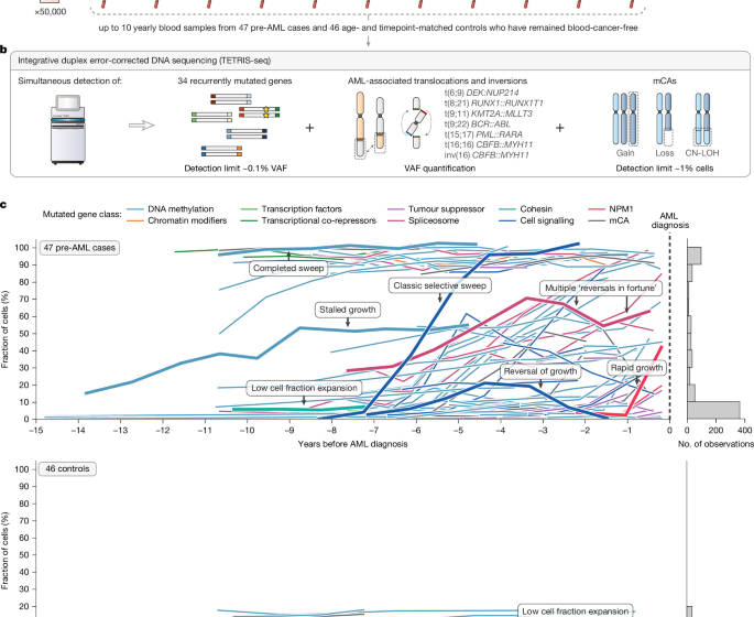
To ensure that we could track the evolution of a comprehensive set of genomic changes associated with clonal hematopoiesis and AML in longitudinal pre-AML and control samples, we developed two separate targeted panels, for use on the same input DNA sample, in an integrated library preparation approach: a small panel (approximately 58 kilobases (kb)) specifically for SNVs and indels and a large panel (approximately 1.6 (megabases (Mb)) for mCA, chromosomal rearrangement and KMT2A partial tandem duplication (KMT2A-PTD) detection (Supplementary Note 2).
For the SNV and indel panel, we designed a custom set of 120 nucleotide (nt) oligonucleotide probes (TWIST Biosciences) targeting commonly mutated exons in 34 genes commonly mutated in clonal hematopoiesis and AML (Supplementary Figures 2–5 and Supplementary Table 1): ASXL1, BCOR, BCORL1, CBL, CEBPA, CHEK2, CSF3R, DDX41, DNMT3A, EZH2, FLT3, GATA2, GNAS, GNB1, HDI1, HDI2, JAK2, KIT, KRAS, MPL, MNP1, NRAS, PPM1D, PTPN11, RAD21, RUNX1, SF3B1, SRSF2, CERF2, TET2, TP53, U2AF1, WT1 And ZRSR2.
For mCA, chromosomal rearrangement and KMT2A-PTD panel, we designed a custom set of 120 nt oligonucleotide probes (TWIST Biosciences) targeting a total of 10,326 common SNPs (minor allele frequency 0.01) spaced approximately every 280 kb across the genome to enable detection of mCAs (Supplementary Table 2). Using this approach, deviations in the B allele frequencies (BAF) of heterozygous SNPs can be detected, which, combined with the depth information read, allows detection of a gain, loss or CN-LOH event. For detection of chromosomal rearrangements, a custom set of 120 nt oligonucleotide baits (TWIST Biosciences) was designed to target known breakpoint regions of each of the rearranged partner chromosomes.61,62,63,64 of seven chromosomal rearrangements that define specific subcategories of AML in the World Health Organization AML classification65:t(6;9) DEK::NUP214t(8;21) RUNX1::RUNX1T1t(9;11) KMT2A::MLLT3t(9;22) RBC::ABLt(15;17) PML::RARAt(16;16) CBFB::MYH11 and inv(16) CBFB::MYH11 (Supplementary Fig. 6 and Supplementary Table 3). KMT2A is noted for having many possible breakpoint partners and thus other common breakpoint regions in KMT2A were also targeted66. KMT2A-PTDs most often involve exon 2 or 3 and span exons 9 to 11 (ref.67). They can be detected by observing a relative increase in reading depth, starting from exon 2 or 3, compared to an exon that is never involved in KMT3A-PTD (e.g. exon 27)68. A custom set of 120 nt oligonucleotide baits…
Gn Health