
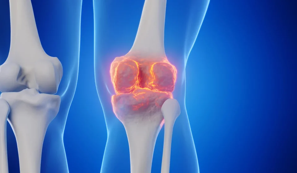
Goodbye joint replacements? Stanford scientists have found a way to regrow cartilage and stop arthritis

Scientists have identified a way to restore cartilage to aging knee joints, at least in mice, by blocking a protein whose levels increase with age. The approach not only reversed the natural loss of cartilage in older animals, but also protected mice from developing arthritis after knee injuries similar to ACL tears in humans.
The results also showed promise in human tissues. Cartilage samples taken during knee replacements responded to treatment by starting to produce new, functional cartilage.
Together, the findings suggest that cartilage damaged by aging or arthritis may be more capable of repairing itself than previously thought. If the strategy ends up working in humans, researchers believe it could potentially lead to an oral drug or injection that regenerates cartilage and reduces the need for knee or hip replacements.
Targeting the damage caused by osteoarthritis
Osteoarthritis is a degenerative joint disease in which cartilage gradually breaks down, leaving joints painful, swollen and increasingly difficult to move. In the United States, it affects approximately one in five adults and is estimated to account for approximately $65 billion in direct health care costs each year.
Current treatments primarily focus on controlling pain and other symptoms. Once the disease becomes severe, surgical replacement of the damaged joint may be the only option left. There are currently no medications that can reliably slow or reverse osteoarthritis itself.
The Stanford Medicine study instead focused on a protein called 15-PGDH. The researchers described it as a gerozyme, a term for enzymes that become more abundant with age and contribute to the progressive loss of tissue function.
Previous work from the same group showed that 15-PGDH acts as an important regulator of aging in several tissues. Blocking the protein with a small molecule increased muscle mass and endurance in old mice. Doing the opposite, increasing 15-PGDH in young animals, caused their muscles to shrink and weaken. The protein has also been linked to the regeneration of bone, nerve and blood cells.
In many of these tissues, healing depends on the multiplication and development of specialized tissue-specific stem cells. Cartilage was found to behave differently. Instead of relying on stem cells, existing cartilage cells called chondrocytes changed their genetic activity patterns and shifted to a younger state.
“This is a new way to regenerate adult tissue, and it shows great clinical promise for treating arthritis caused by aging or injury,” said Helen Blau, PhD, professor of microbiology and immunology. “We were looking for stem cells, but they are clearly not involved. It’s very exciting.”
Blau, who directs the Baxter Laboratory for Stem Cell Biology and is a Donald E. and Delia B. Baxter Foundation Professor, and Nidhi Bhutani, PhD, associate professor of orthopedic surgery, are senior authors of the research, published in Science. Instructor of orthopedic surgery Mamta Singla, PhD, and former postdoctoral researcher Yu Xin (Will) Wang, PhD, are lead authors of the study. Wang is now an assistant professor at the Sanford Burnham Institute in San Diego.
“Spectacular regeneration” of cartilage
“Millions of people suffer from joint pain and swelling as they age,” Bhutani said. “This is a huge unmet medical need. Until now, no drug has directly addressed the cause of cartilage loss. But this gerozyme inhibitor causes dramatic cartilage regeneration beyond that reported in response to any other drug or intervention.”
Cartilage is not the same at all. The human body contains three main types, each designed for a different job.
Elastic cartilage is soft and flexible and helps form structures such as the outer ear. Fibrocartilage is stronger and better suited to absorb force, including in the areas between the vertebrae of the spine. Hyaline cartilage is smooth and slippery, allowing bones to move against each other with very little friction in joints such as the ankles, hips, shoulders and parts of the knee.
In joints, hyaline cartilage is also called articular cartilage. This is the type most often damaged by osteoarthritis.
Osteoarthritis can develop when joints experience stress from aging, injury or obesity. Chondrocytes begin to produce inflammatory molecules while breaking down collagen, a structural protein that gives cartilage much of its strength.
As collagen disappears, cartilage becomes thinner and softer. The inflammation adds swelling and pain, creating the familiar symptoms of osteoarthritis.
The problem is that articular cartilage normally has very little ability to repair itself. Researchers have identified possible stem or progenitor cells capable of forming cartilage in bone, but efforts to find similar cell populations within articular cartilage itself have not been successful.
An aging protein becomes a target
Previous research from Blau’s lab found that a molecule called prostaglandin E2 is important for muscle stem cell function. The 15-PGDH protein breaks down prostaglandin E2.
Blocking 15-PGDH, or increasing prostaglandin E2 levels, has been shown to promote the regeneration of damaged muscles, nerves, bones, colon, liver and blood cells in young mice.
This raised an important question for Blau, Bhutani and their colleagues: Could the same biological pathway contribute to cartilage deterioration as animals age or recover from injury?
The researchers compared the levels of 15-PGDH in the knee cartilage of young and old mice. They found that the amount of gerozyme roughly doubled with age.
They then gave the older animals a small molecule drug designed to inhibit 15-PGDH. In one experiment, the drug was injected into the abdomen in order to have effects throughout the body. In another case, it was administered directly into the knee joint.
Both approaches produced striking results.
The cartilage in the knees of the older animals was noticeably thinner and less functional than that of the young mice. However, after treatment, it became thicker over the entire joint surface.
Importantly, the cells produced hyaline cartilage, the smooth articular cartilage needed for healthy joint movement, rather than less suitable fibrocartilage.
“Such cartilage regeneration in aged mice surprised us,” Bhutani said. “The effect was remarkable.”
Protecting Knees After ACL Injuries
The researchers also tested whether the treatment could help after traumatic knee injuries.
ACL tears are especially common in sports like football, basketball, and skiing, where athletes pivot, stop abruptly, or jump frequently. Surgery can repair the torn ligament, but repairing the immediate injury does not necessarily prevent long-term joint damage.
About 50% of people who sustain these injuries develop osteoarthritis in the affected joint within about 15 years.
In the mouse experiments, researchers administered the gerozyme inhibitor twice a week for four weeks after injury. The treatment significantly reduced the risk of the animals developing osteoarthritis.
Mice given a control drug had twice the levels of 15-PGDH compared to animals whose knees had not been injured and developed osteoarthritis within four weeks.
The treated mice also moved more normally and put more weight on the paw attached to the injured leg than the untreated animals.
“Interestingly, prostaglandin E2 has been implicated in inflammation and pain,” Blau said. “But this research shows that at normal biological levels, small increases in prostaglandin E2 can promote regeneration.”
Make old cartilage cells younger
To understand what was happening inside the joint, the researchers took a closer look at chondrocytes from young and old mice.
Older cartilage cells showed more activity in genes associated with inflammation and unwanted conversion of hyaline cartilage to bone. At the same time, genes involved in normal cartilage development were less active.
The treatment changed this balance.
A population of old chondrocytes producing 15-PGDH and expressing genes associated with cartilage breakdown decreased from 8% of cells to 3%.
A second population, which did not produce 15-PGDH but expressed genes related to fibrocartilage formation, dropped from 16% to 8%.
Meanwhile, a third group was moving abruptly in the opposite direction. These cells did not produce 15-PGDH and expressed genes involved in the formation of hyaline cartilage and in the maintenance of the extracellular matrix necessary for the proper functioning of cartilage. Their share increased from 22% to 42%.
The extracellular matrix is the network of proteins and other molecules that surrounds cells and gives tissues their structure. In cartilage, this is especially important because it helps the tissues resist pressure while maintaining the smooth joint surface they need for movement.
Overall, the treatment appears to push cartilage toward a younger biological state without recruiting stem or progenitor cells.
Human cartilage also responded
The researchers then looked at cartilage taken from people with osteoarthritis who were undergoing total knee replacements.
After one week of treatment with the 15-PGDH inhibitor, human tissue contained fewer chondrocytes expressing 15-PGDH. The activity of genes associated with cartilage and fibrocartilage degradation was also decreased compared to untreated tissues.
Most notably, the samples began to regenerate articular cartilage.
“The mechanism is quite striking and has really changed our perspective on how tissue regeneration can occur,” Bhutani said. “It is clear that a large number of cells already existing in cartilage are changing their gene expression patterns. And by targeting these cells for regeneration, we may have the opportunity to have a greater overall clinical impact.”
The results do not yet establish that the treatment can regrow cartilage or prevent osteoarthritis in humans. The mouse results…
Gn Health