

Clinical cohorts
Ethics Statement
The UCLH bronchoscopy study and the SUMMIT, ALPINE and Molecular Pathogenesis of Lung Disease II studies have been approved by the UK Health Research Authority (HRA). The ASCENT study was approved by the HRA and Health and Care Research Wales. All human participants provided written informed consent prior to enrollment. The animal studies were approved by the University College London Biological Services Review Committee and were carried out in accordance with the UK Animals (Scientific Procedures) Act 1986 and associated Home Office guidelines.
UCLH Bronchoscopy Surveillance Study Cohort
The UCLH Bronchoscopy Surveillance Study16.18 (REC: 01/0148) is a longitudinal study of patients with preinvasive lesions of the bronchus. It aims to determine the nature of bronchial lesions and to analyze the molecular, histological and immunocytochemical modifications associated with their evolution towards invasion. Included patients must be able to provide informed written consent to participate, have pre-invasive lesions of the bronchus, and be 18 years of age or older. Patients were excluded from the study if they were unable or unwilling to provide informed consent, had a diagnosis of invasive lung carcinoma at the time of enrollment, specific respiratory diseases or other medical conditions affecting tolerance of bronchoscopy, or had coagulation abnormalities.
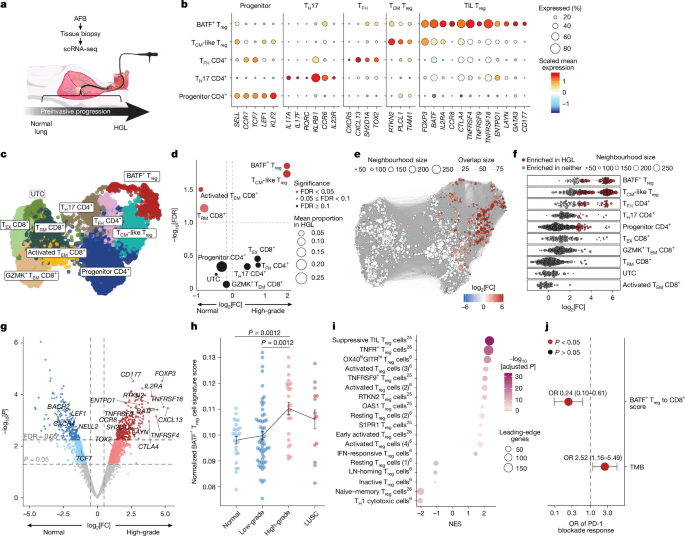
This surveillance study targeted individuals at high risk of lung cancer (i.e., heavy smokers) and uses BFA to screen for preinvasive lesions in the bronchus. Preinvasive lesions of the central airways are classified according to a severity spectrum based on their histology. Low-grade lesions include hyperplasia, metaplasia, mild dysplasia, and moderate dysplasia. HGLs include severe dysplasia, CIS, and microinvasive CIS (CIS+). Patients with low-grade lesions were monitored by AFB approximately once a year and patients with HGL were monitored every 3 to 6 months. For annotation of lesion grade for peripheral blood samples, the highest grade lesion identified at the corresponding bronchoscopy visit was assigned. For these analyses, patients with inconsistent lesion grade over time were excluded from the cross-sectional analysis.
SUMMIT and ASCENT cohorts
The ASCENT study (REC: 20/SC/0128, NCT04204499)32 is a prospective, observational cohort study of participants undergoing surgical resection for lung cancer detected by low-dose computed tomography (LDCT). Participants will have undergone LDCT screening in the SUMMIT study (REC: 17/LO/2004, NCT03934866)34. SUMMIT is a prospective, observational cohort study that aims to evaluate the implementation of LDCT lung cancer screening in a diverse, high-risk population in London and to validate an early detection blood test for multiple cancers. People aged 55 to 77 years who had been recorded as current smokers and who were not on the palliative care register of their National Health Service (NHS) England primary care records at any time in the previous 20 years were identified to be invited for a lung health check. Individuals who met the 2013 USPSTF criteria or had a 2012 prostate, lung, colorectal, and ovarian (PLCO) patternM2012) A 6-year risk ≥1.3% and not currently receiving treatment for active cancer (except adjuvant hormonal therapy) was asked to provide informed consent to participate in the study. Blood samples were collected at surveillance time points before cancer diagnosis (SUMMIT), and matched blood, tumor, and adjacent histologically normal tissue samples were collected at the time of surgical resection (ASCENT). Patients were excluded from the ASCENT study if they had active infection with HIV, hepatitis B virus, hepatitis C virus, or syphilis, or if the local multidisciplinary team decided to treat the current lung malignancy with neoadjuvant therapy.
The NSCLC samples in this study include both LUAD and LUSC samples at stages I to III, with 90% of the samples analyzed being classified as stage I. Upon resection, some suspected NSCLC cases were found to be noncancerous and therefore were excluded from the analyses. These samples were classified as benign inflammations or tumors and included granulomas, benign tumors, chronic inflammation, smoking-related changes, scarring, necrotizing granulomatous inflammation, non-necrotizing granulomatous inflammation, fungal balls, dendriform lung ossification, or lymph node changes.
Blood samples from healthy, age-matched controls were collected from patients at the UCLH Orthopedic Pre-Assessment Clinic prior to hip replacement surgery. Patients included were aged 50 years or older and were able to provide written informed consent. Patients with active infections, history of malignancy, autoimmune diseases, or abnormal blood counts were excluded. For flow cytometry experiments, we also included samples from healthy donors obtained from cone leukocytes from NHS blood and transplantation residual platelet donation.
ALPINE cohort
Biomarkers in lung health checks (ALPINE)33: Evaluation of Novel Biomarkers in Participants Undergoing Targeted Lung Health Checks (REC: 23/EM/0114, NCT05902559) is a prospective, observational cohort study of minimally invasive samples from patients undergoing screening in the North Central London Targeted Lung Health Check program. The Targeted Lung Health Check program is an NHS program which aims to use LDCT screening in people at high risk of lung cancer to improve early diagnosis and survival. It is also a continuation of SUMMIT, which enables collection of blood and PBMC for immune analysis and helps with recruitment to the ASCENT study once participants develop NSCLC and undergo surgical resection. Participants must be aged 55 to 74 and be smokers or former smokers. In this study, whole blood and PBMC samples from the ALPINE study were used as “cancer-free” controls, selecting samples from participants undergoing LDCT screening but without any nodules presented on imaging at the time of sampling.
Molecular pathogenesis of cohort II lung diseases
An investigation into the molecular pathogenesis of lung disease II (REC: 06/Q0505/12, IRAS: 245471)57 is a prospective observational cohort established to study molecular changes in human airway epithelium associated with the development and progression of lung disease. Participants are recruited from individuals undergoing a range of respiratory diagnostic and clinical procedures, including bronchoscopy, upper respiratory endoscopy, thoracic surgery (including video-assisted thoracoscopic surgery and surgical resection), radiology-guided biopsy, outpatient respiratory clinics or inpatient admissions. Eligible participants are adults (≥18 years) under investigation for respiratory symptoms or being monitored for respiratory illness. For this study, only tumors and corresponding adjacent parenchyma from participants with a confirmed diagnosis of lung cancer were included.
For the ASCENT and Molecular Pathogenesis of Lung Disease II cohorts, lung tissue was defined pathologically using the UICC TNM classification, eighth edition, of malignancies by pathologists.
Collection and profiling of human samples
Processing tissue samples for single cell sequencing
Tissue biopsy samples from lesions collected during AFB (Supplementary Table 1) were finely cut using sterile scalpel blades and digested in dissociation buffer containing 100 µg ml.–1 DNase I (Sigma, DN25) and 1 mg ml–1 collagenase (Sigma, C9407) in HBSS (Gibco, 14025092) at 37 °C for 1 h with continuous shaking. Biopsy suspensions were filtered through a 70 µm cell strainer and washed once in PBS with 10% BSA, followed by incubation in 1× red blood cell (RBC) lysis buffer (Miltenyi Biotec, 130-094-183) for 2 min at room temperature (RT). Single-cell suspensions were then prepared for 10x sequencing by washing twice in PBS with 0.04% BSA. The cell concentration was adjusted to 1,000 cells per µl and samples were processed either with a Chromium Next GEM Single Cell 5′ v.2 kit (10x Genomics, 1000265), with or without TCR amplification, according to the manufacturer’s instructions. The libraries were sent to Novogene for sequencing.
For two bronchial biopsy samples with replicate technical preparations, P166 normal and P168 LT CIS, the replicate libraries were pooled before downstream analyzes and treated as a single biological sample for all sample-level analyses.
Blood sample processing
Blood samples were collected in Vacutainer EDTA blood collection tubes (BD Biosciences, 366643). PBMCs were isolated from whole blood by density gradient centrifugation (750g10 min, TA, without brake) with Ficoll Paque Plus (Cytiva, 17144003). The lymphocyte interface was washed twice with complete RPMI 1640 (Merck, R0883), resuspended in a solution containing FBS (PAN Biotech, P40-37500) supplemented with 10% DMSO (Merck, D2650-100ML), and cryopreserved in liquid nitrogen before downstream use. When the buffy coats were also collected, the Vacutainer EDTA tubes were centrifuged (1,800g15 min, RT, no brake) and the buffy coat interface was frozen directly in cryogenic vials at −80 °C and stored until downstream use.
Preparation of PBMC samples for single-cell sequencing
Cryopreserved PBMC samples were thawed using warm R20 medium (RPMI 1640 with 20% FBS, 2 mM I-glutamine (Merck, G7513-100ML), 10 mM HEPES (ThermoFisher Scientific, 15630056) and 100 U ml–1 penicillin–streptomycin (Merck, P0781-100ML)) and washed with R10 medium (RPMI 1640 with 10% FBS and 100 U ml–1 penicillin–streptomycin) containing 37.5 µg ml–1 DNase I. Cells were washed twice in FACS buffer.
For matched PBMC samples from the UCLH bronchoscopy surveillance cohort, non-naïve T cell sorting was performed. Cells were stained with CCR7 and Fc receptor binding inhibitor polyclonal antibody (Invitrogen, 14-9161-73) for 15 minutes at 37°C. Then, an additional 30 minutes of incubation at 4°C was carried out with the…
Gn Health